


By María I Gaitán, MD1, Paulina Yanez, MD2 and Eduardo H.M.S G Figueiredo BSc3
1Staff Clinician, Neuroimmunology Section, Neurology Department, FLENI, Buenos Aires,Argentina.
2Neuroradiologist, Vice-Head Department of Diagnostic Imaging , FLENI, Buenos Aires, Argentina.
3MR CML & Research, GE Healthcare LATAM, Sao Paulo, Brazil.
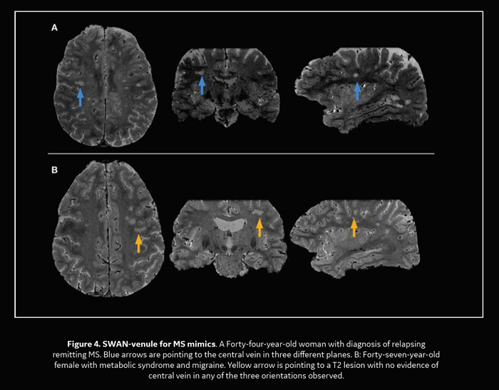
“The central vein sign (CVS), defined as the hypointense area in the center of MS lesions seen as a dot or a line, is very specific to distinguish MS from its radiological mimics such as AQP4-IgG-positive NMOSD, systemic autoimmune diseases, cerebral small vessels diseases, Susac syndrome, and white matter migraine lesions. By optimization of the conventional SWAN sequence we were able to provide a sensitive technique for the detection of the CVS at 3.0T in a clinical setting, calling SWAN-venule.”
About the authors from FLENI:


Contributors GE:
Thomas Doring, PhD, Collaborations GROE, GE Healthcare Latam. Eduardo Figueiredo, CMLResearch MR Latam.
FLENI is a non-profit foundation created in 1959, with the initial objective of contributing to the prevention and fight against neurological diseases of childhood. The Magnetic Resonance service is specialized in studies of the central and peripheric nervous system. The MRI service has two 3T Discovery 750 scanners from GE Healthcare that were, recently updated to the newest software release DV26.
Introduction
Multiple sclerosis (MS) is the most common neurological disorder in young adults. Conventional Magnetic Resonance Imaging (MRI) is very sensitive to establish MS dissemination in time and space but not as specific to discriminate MS from other white matter diseases. Pathological studies from last century have shown that MS lesions develop around small veins and venules, being a pathological biomarker of the disease.
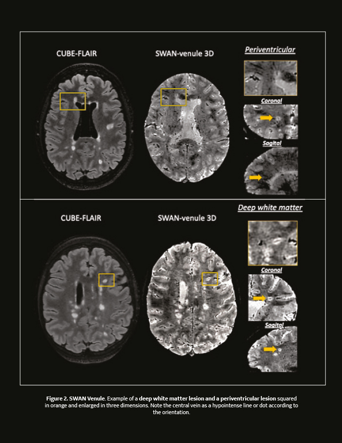
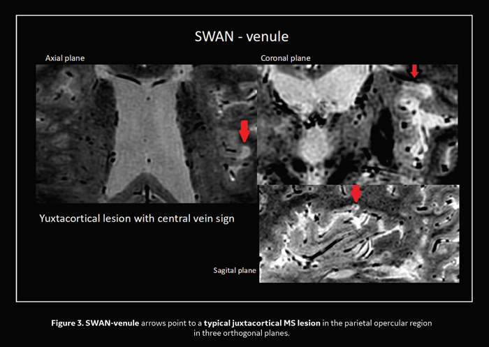
With high resolution, susceptibility-based MRI (e.g. T2*-weighted, susceptibility weighted imaging) and base on the paramagnetic nature of deoxyhemoglobin, these veins can be visualized centrally at 7.0 T MRI within most (80-100%) MS lesions.1 Moreover, recently the North American Imaging in Multiple Sclerosis cooperative (NAIMS), proposed the radiological definition of the central vein sign (CVS) as the hypointense area in the center of MS lesions seen as a dot or a line.
Interestingly, CVS is very specific to distinguish MS from its radiological mimics such as AQP4-IgGpositive NMOSD, systemic autoimmune diseases, cerebral small vessels diseases, Susac syndrome, and white matter migraine lesions.
At 3.0 Tesla, a T2*weighted 3D echo-planar-imaging sequence, has shown higher detection of the CVS than standard susceptibility-weighted Imaging (SWAN), which is not sensitive enough to detect the CVS.3 However, it is possible to properly optimize the conventional SWAN sequence protocol to provide a sensitive technique for the detection of the CVS at 3.0T in a clinical setting, calling SWAN-venule4.
The purpose of this report is to share our experience in using SWAN-venule in our 3.0 Tesla (Discovery MR750, GE Healthcare, MKE, USA).
MR Protocol
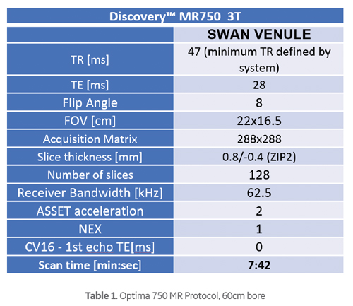
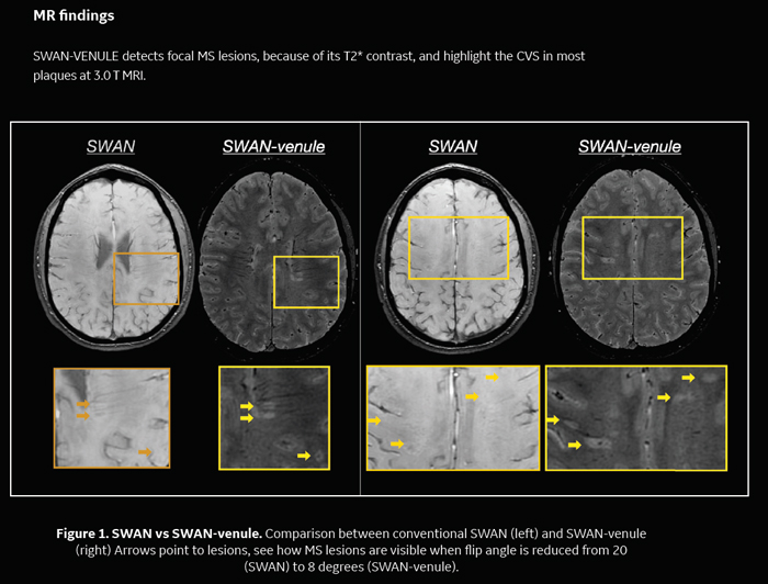
The goal was optimizing the SWAN sequence, commercially available for a long time, mainly by reducing the Flip Angle to 8 degrees, thus more PD to T2* weighting, reducing T1 weighting, and adapting it to 0.8mm isometric spatial resolution, to be able to reformat in any imaging plane maintaining same resolution. Additionally, in order to improve sensitivity to the detection of veins, instead of increasing the echo time to more than 28.5ms, the sequence is acquired post gadolinium injection, as this increases susceptibility eff ects inside the vessel.
The protocol can be easily adapted to any 3.0 Tesla MRI equipment that has the SWAN sequence license. One characteristic of the MR750 3.0 Tesla scanner is its high gradient performance, enabling very short TR and with it reduced acquisition times. When someone has the option to choose between the two coils, the 32ch head coil is the preferred one over HNS Head coil due to its improved SNR. SWAN is the commercial name of the generic susceptibility weighted pulse sequence, which protocol can be adjusted to result in more T1, PD or T2* weighting.
SWAN-venule was a given name to reflect the methodology and Central Vein Sign detection. We apply this modified SWAN protocol in the brain MRI protocol for demyelinating diseases and we add it in patients with other disorders that simulate MS in the clinical practice.
Key points
• SWAN-venule is sensitive for detection of central veins inside brain lesions at clinical 3T.
• This modified-optimized sequence protocol is recommended to be included in brain MRI protocols for demyelinating diseases in the clinical practice. This sequence and protocol are available on most systems.
• Radiologists and neurologists can assess the CVS and use it as a biomarker to discriminate MS from its radiological mimics.
References
1-Tallantyre EC, Brookes MJ, Dixon JE, Morgan PS, Evangelou N, Morris PG. Demonstrating the perivascular distribution of MS lesions in vivo with 7-Tesla MRI. Neurology. 2008;70(22):2076-2078.
2-Sati, P., Oh, J., Constable, R. et al. The central vein sign and its clinical evaluation for the diagnosis multiple sclerosis: a consensus statement from the North American Imaging in Multiple Sclerosis Cooperative. Nat Rev Neurol 12, 714–722 (2016).
3-Samaraweera AP, Clarke MA, Whitehead A, et al. The Central Vein Sign in Multiple Sclerosis Lesions Is Present Irrespective of the T2* Sequence at 3 T. J Neuroimaging. 2017;27(1):114-121.
4-Gaitán MI, P. Yañez, M.E. Paday Formenti, I. Calandri, E. Figueiredo, P. Sati and J. Correale SWAN-Venule: An Optimized MRI Technique to Detect the Central Vein Sign in MS plaques M. .American Journal of Neuroradiology February 2020, DOI:https://doi.org/10.3174/ajnr.A6437


